How often do we truly appreciate our eyes and what they do for us? Imagine your eyes as a biological camera, meticulously capturing snapshots of the world and transmitting them to your brain for processing and interpretation. According to Medical News & Life Sciences, humans have binocular vision, meaning that both the eyes create a single combined image. Optical components create an image, which further gets perceived and interpreted by the brain via connecting neurons. The entire machinery works in quite an intricate manner.
Why Eye Anatomy Matters
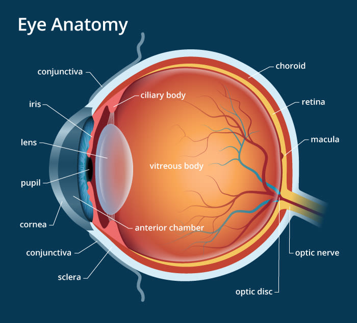
The human eye is a complex system where every component plays a critical role in delivering clear vision. Understanding its parts helps you appreciate how vision works and why conditions like nearsightedness or astigmatism occur. According to Medical News Today, humans rely on binocular vision, where both eyes combine to form a single image, processed by the brain through intricate neural pathways. Below, we break down the eye’s anatomy and its impact on your sight.
Key Parts of the Human Eye
Each part of the eye has a unique function, working together to transform light into the images you see. Here’s a detailed look at the eye’s anatomy:
1. Orbit: The Protective Socket
The orbit is the bony cavity in your skull that houses the eye. It contains six extraocular muscles that attach to the eye, enabling movements like looking side-to-side, up and down, or rotating. This structure protects the eye while allowing precise control.
- Function: Protects the eye and supports movement.
- Vision Impact: Ensures smooth eye movements for tracking objects.
2. Sclera: The Eye’s Outer Shield
The sclera is the white, tough outer layer of the eyeball. Covering most of the eye’s surface, it provides structural support and serves as an attachment point for the extraocular muscles.
- Function: Maintains eye shape and strength.
- Vision Impact: Stabilizes the eye for consistent vision.
3. Conjunctiva: The Moisture Barrier
This thin, transparent tissue covers the front of the eye and lines the inside of the eyelid. When blood vessels in the conjunctiva enlarge, your eyes may appear red, often due to irritation or allergies.
- Function: Keeps the eye moist and protects it from irritants.
- Vision Impact: Prevents discomfort that could blur vision.
4. Cornea: The Focusing Lens
The cornea is the clear, dome-shaped surface at the front of the eye. It bends light to focus it onto the retina, playing a key role in sharp vision. Changes in its curvature can lead to blurry vision, like in astigmatism.
- Function: Focuses light for clear images.
- Vision Impact: Curvature issues (like astigmatism) can cause blurry vision.
5. Lens: The Adjustable Focus
Located behind the cornea, the lens fine-tunes light focus onto the retina. It changes shape—thicker for near objects, thinner for distant ones—to ensure clarity at varying distances.
- Function: Adjusts focus for near and far vision.
- Vision Impact: Enables clear vision at varying distances.
6. Ciliary Muscles: The Lens Adjusters
These tiny muscles surround the lens, contracting or relaxing to adjust its shape. For close-up tasks, they contract to thicken the lens; for faraway objects, they relax to thin it.
- Function: Adjusts lens shape for focusing.
- Vision Impact: Ensures sharp focus for reading or viewing far objects.
7. Retina: The Light-Sensing Layer
The retina, at the back of the eye, contains rods and cones—cells that detect light and color. It converts light into electrical signals, forming the images you see.
- Function: Detects light and forms images.
- Vision Impact: Enables color perception and visual clarity.
8. Macula: The Center of Sharp Vision
Found within the retina, the macula is responsible for central vision and fine details, like reading or recognizing faces. Damage to it can impair these abilities.
- Function: Provides sharp, central vision.
- Vision Impact: Damage can impair tasks like reading or driving.
9. Optic Nerve: The Brain’s Messenger
The optic nerve carries electrical signals from the retina to the brain, where they’re interpreted as visual images. It’s the final step in the vision process.
- Function: Transmits visual data to the brain.
- Vision Impact: Essential for processing what you see.
10. Vitreous Humor: The Eye’s Cushion
This clear, jelly-like substance fills the space between the lens and retina. It maintains the eye’s shape and protects internal structures.
- Function: Supports eye shape and cushions internal parts.
- Vision Impact: Ensures structural stability for clear vision.
11. Iris: The Colorful Controller
The iris, the colored part of your eye, regulates light entry by adjusting the pupil’s size. Its unique pigmentation is inherited genetically.
- Function: Controls light entering the eye.
- Vision Impact: Optimizes vision in bright or dim conditions.
12. Pupil: The Light Gateway
The pupil, the black center of the iris, controls light intake. It widens in dim light (up to 7 mm) and shrinks in bright light (down to 3 mm) to optimize vision.
- Function: Regulates light entry.
- Vision Impact: Enhances vision clarity in varying lighting.
13. Eyelids: The Protective Curtains
Eyelids shield the eye from light during sleep and blink instinctively to keep the eye moist and free of debris.
- Function: Shields and lubricates the eye.
- Vision Impact: Prevents dryness or irritation that could blur vision.
14. Tear Glands: The Moisture Makers
Located in the upper eyelid, these glands produce tears to lubricate and clean the eye’s surface, preventing damage from dryness.
- Function: Produces tears to maintain eye health.
- Vision Impact: Prevents dryness for clear, comfortable vision.
15. Eyelashes and Eyebrows: The Dust Blockers
These hairs trap dust, sweat, and debris, protecting the eye from irritation and maintaining clear vision.
- Function: Protects against foreign particles.
- Vision Impact: Maintains clear vision by reducing irritation.
| Eye Part | Function | Impact on Vision |
|---|---|---|
| Cornea | Focuses light onto retina | Affects clarity; curvature issues cause blurriness |
| Lens | Adjusts focus for near/distant objects | Ensures sharp images at varying distances |
| Retina | Converts light to electrical signals | Enables color and light perception |
| Optic Nerve | Transmits signals to brain | Delivers visual information for processing |
| Iris/Pupil | Regulates light entry | Optimizes vision in different lighting |
How Vision Problems Arise
When any part of the eye doesn’t function properly, vision issues can occur. For example:
- Cornea curvature changes (e.g., astigmatism) distort light focus, causing blurry vision.
- Lens flexibility loss (e.g., presbyopia) makes focusing on close objects difficult.
- Retina issues (e.g., macular degeneration) impair central vision or color perception.
Corrective lenses, like glasses or contacts, adjust light entry to compensate. However, many people opt for LASIK eye surgery for a permanent solution.
LASIK: A Path to Clearer Vision
Tired of foggy glasses or uncomfortable contacts? LASIK eye surgery reshapes the cornea to correct vision issues like nearsightedness, farsightedness, or astigmatism. Benefits include:
- Long-term clarity: Most patients achieve 20/20 vision or better.
- Quick recovery: Return to normal activities within days.
- Freedom from lenses: No more glasses or contacts for most.
Pro Tip: Schedule a free consultation to discuss your vision goals with a LASIK specialist. Call 866.229.4570 or find a location near you!
Is LASIK Right for You?
Eligibility depends on factors like eye health, corneal thickness, and prescription stability. Consult a qualified eye care professional to assess your needs. At The LASIK Vision Institute, our experts use advanced technology to personalize your treatment.
Frequently Asked Questions

Take the Next Step Toward Better Vision
Your eyes work tirelessly to bring the world into focus. Understanding their anatomy empowers you to protect your vision and explore options like LASIK for lasting clarity. Ready to ditch glasses or contacts? Contact The LASIK Vision Institute or schedule your free consultation today. Start your journey to sharper vision now!

Find a LASIK Surgery Location Near You
We’re located nationwide – it’s easy to find a LASIK Vision Center near you.
Categories: